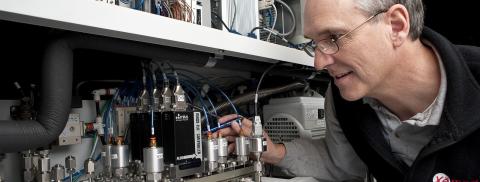
UNH physicists are engaged in fundamental research in producing nuclear polarization in gases and applying those techniques for medical imaging applications. Lung disease is the fourth leading cause of death in the US, yet there is no widely available modality to noninvasively image lung structure and function. Hyperpolarized gases, noble gases with their magnetic polarization enhanced by a factor of ten thousand, can be harmlessly breathed and imaged in the lungs. Imaging techniques can be devised to reveal gas space structure as well as functional information such as ventilation maps, local microstructure, and oxygen uptake.
UNH presently leads the world by more than a factor of ten in the quality and quantity of hyperpolarized xenon production. We are collaborating with imaging scientists at the Brigham and Women's Hospital in Boston to apply this technology to measuring lung surface-to-volume ratio in animals and humans. At our Center for Hyperpolarized Gas Studies we are implementing precision diagnostics to quantify the rate and degree of polarization, in particular by measuring the EPR shift of the rubidium fine structure due to polarized xenon, and we are investigating whether hyperpolarized xenon may be useful for providing early diagnosis of other diseases, such as cancer.
Applied Optics Group
Over the past twenty years, members of the UNH Physics Department have been investigating Spin Exchange Optical Pumping (SEOP) to identify new technologies for producing nuclear polarized gases. Originally these efforts were motivated by applications in fundamental physics. Because the lone neutron in 3He dominates the spin and magnetic properties of the composite nucleus, a dense gas of highly polarized 3He can serve as a surrogate neutron target for electron scattering experiments. Also, polarized3He preferentially absorbs neutrons of opposite spin direction allowing volumes of polarized 3He gas to serve as analyzers of neutron polarization in neutron scattering experiments.
More recently our efforts to improve SEOP technologies are also motivated by opportunities to apply hyperpolarized gas as a diagnostic tracer of inhaled lung gas with Magnetic Resonance Imaging. Typically an MRI scanner will study the tissues of the human body by aligning (polarizing) the protons in these tissues using its strong magnetic field. However SEOP can achieve hyperpolarizations nearly a million times greater. Since 1997 our group has been pursuing a new method for hyperpolarizing 129Xe for medical imaging. Our method flows axenon gas mixture at low pressure and high velocity through a long chamber against the direction of propagation of the laser beam, efficiently accumulating very high polarizations. This technology motivated the spinout of a small startup company Xemed LLC.
The UNH Center for Hyperpolarized Nuclei collaborates with Xemed and with clinical partners nationwide to demonstrate applications of hyperpolarized 129Xe. Recent UNH Physics graduate student Isabel Dregely performed Ph.D. thesis work by collaborating with Xemed and outside academic institutions. In collaboration with the Martinos Center for Biomedical Imaging at the Massachusetts General Hospital we implemented a 32-elementchest coil for accelerated parallel imaging. In collaboration with the University of Virginia Center for In-Vivo Hyperpolarized Gas MR Imaging we implemented new scanner pulse-sequences to interrogate the saturation rate of xenon entering lung tissues. For her work, Isabel Dregely was honored with the W.S Moore Young Investigator Award by the International Society of Magnetic Resonance in Medicine.
Current projects include refinements of the technology for polarizing helium-3 and xenon-129 and clinical applications. Our technical projects include polarizer refinements to improve polarization and reduce losses, methods to move polarized gases both locally and over long distances without losing the polarization, development of new powerful line-narrowed pump laser architectures, and development of an inhalation apparatus and RF coil to image premature infants. Clinical studies with collaborators include investigating methods to image premature infants (with University of Missouri) and guiding bronchial thermoplasty to modify airways of severe asthmatics (with Washington University in St. Louis).
Professor: Bill Hersman